The Anatomy and Function of the Human Elbow
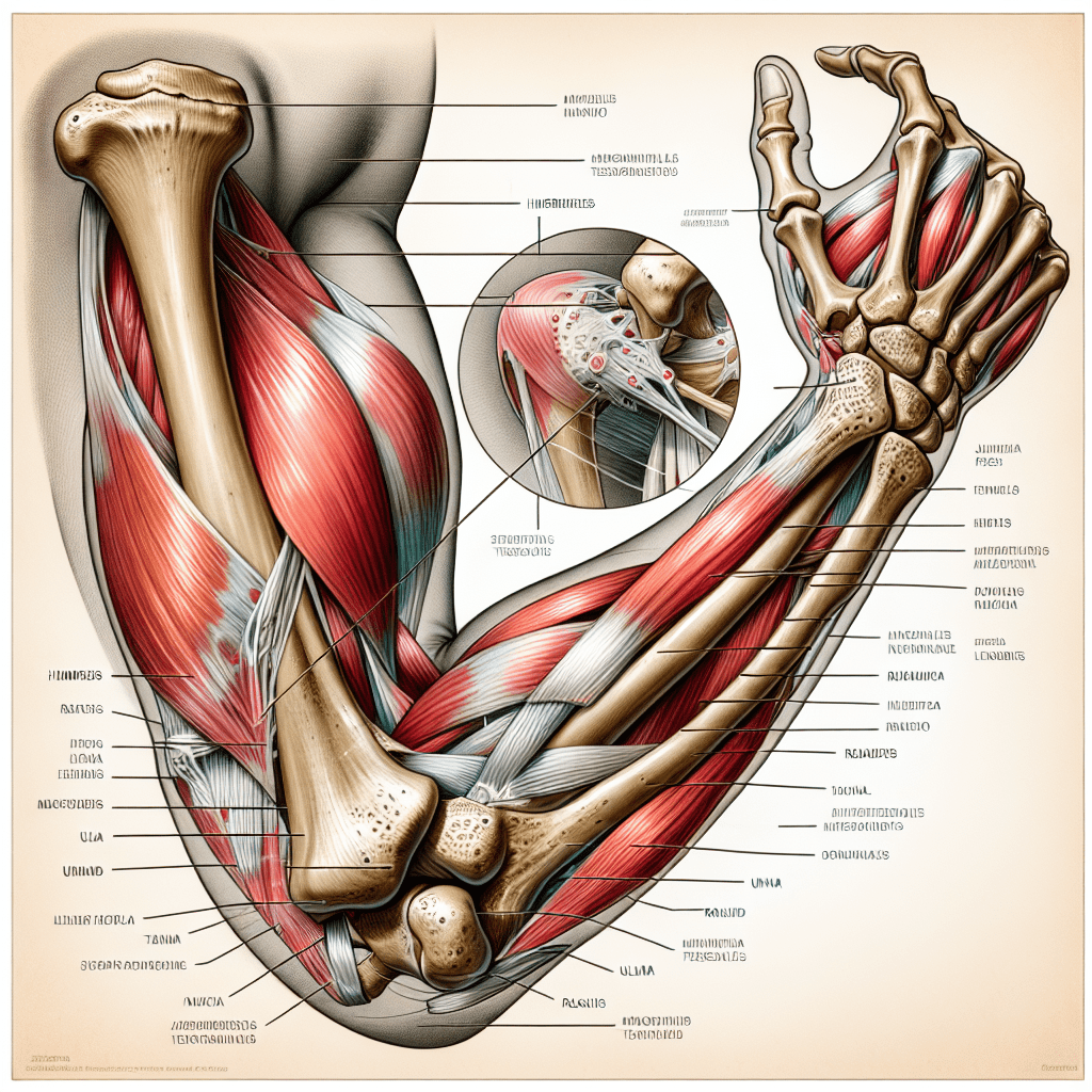
The elbow is an intricate and important joint in the human body that plays a crucial role in arm movement and dexterity. Situated at the juncture where the upper arm bone (humerus) meets the two bones of the forearm (radius and ulna), the elbow enables hinge-like motions along with a rotation of the forearm, making it essential for tasks like lifting, throwing, and many more. Understanding the anatomy and function of the elbow can shed light on how this joint works, how it can become injured, and how those injuries are treated.
Structural Components of the Elbow
The elbow joint is composed of three bones, supported by muscles, tendons, and ligaments that provide stability and facilitate movement.
–
Bony Anatomy : The bottom end of the humerus features two protuberances called the medial and lateral epicondyles. The ulna, when extended, forms the prominent point of the elbow known as the olecranon. The radius allows for forearm rotation.
– Ligaments : The medial (ulnar) collateral ligament, lateral (radial) collateral ligament, and annular ligament stabilize the elbow against various stresses.
– Muscles and Tendons : Major muscles involved include the biceps brachii, brachialis, triceps brachii, and various forearm muscles. Tendons attach these muscles to bone; notably, the biceps tendon connects to the radius, allowing for powerful flexion. Elbow Joint Mechanics
–
Ligaments : The medial (ulnar) collateral ligament, lateral (radial) collateral ligament, and annular ligament stabilize the elbow against various stresses.
– Muscles and Tendons : Major muscles involved include the biceps brachii, brachialis, triceps brachii, and various forearm muscles. Tendons attach these muscles to bone; notably, the biceps tendon connects to the radius, allowing for powerful flexion. Elbow Joint Mechanics
–
Muscles and Tendons : Major muscles involved include the biceps brachii, brachialis, triceps brachii, and various forearm muscles. Tendons attach these muscles to bone; notably, the biceps tendon connects to the radius, allowing for powerful flexion. Elbow Joint Mechanics
Elbow Joint Mechanics
Governing much of our hand manipulation capabilities, elbow joint mechanics feature two key types of movements:
–
Flexion and Extension : Hinging movements conducted in the sagittal plane wherein flexion brings the hand toward the shoulder while extension straightens the arm.
– Pronation and Supination : Rotational movements carried out by the radius and ulna allowing the palm to turn face down or up respectively. Common Elbow Injuries and Disorders
–
Pronation and Supination : Rotational movements carried out by the radius and ulna allowing the palm to turn face down or up respectively. Common Elbow Injuries and Disorders
Common Elbow Injuries and Disorders
The elbow can be susceptible to various injuries due to trauma, overuse, or degeneration:
–
Tennis Elbow : A form of tendinitis commonly resulting from repetitive motions that lead to pain on the outside of the elbow.
– Golfer’s Elbow : Similar to tennis elbow but affects the inside of the joint.
– Elbow Fractures : Trauma often leads to breaks in one or more bones forming the joint.
– Dislocations : An injury where bones slip out of place, frequently caused by falls or impacts.
– Osteoarthritis : Wear-and-tear arthritis that may develop over time causing pain and stiffness. Rehabilitation and Treatment Strategies
–
Golfer’s Elbow : Similar to tennis elbow but affects the inside of the joint.
– Elbow Fractures : Trauma often leads to breaks in one or more bones forming the joint.
– Dislocations : An injury where bones slip out of place, frequently caused by falls or impacts.
– Osteoarthritis : Wear-and-tear arthritis that may develop over time causing pain and stiffness. Rehabilitation and Treatment Strategies
–
Elbow Fractures : Trauma often leads to breaks in one or more bones forming the joint.
– Dislocations : An injury where bones slip out of place, frequently caused by falls or impacts.
– Osteoarthritis : Wear-and-tear arthritis that may develop over time causing pain and stiffness. Rehabilitation and Treatment Strategies
–
Dislocations : An injury where bones slip out of place, frequently caused by falls or impacts.
– Osteoarthritis : Wear-and-tear arthritis that may develop over time causing pain and stiffness. Rehabilitation and Treatment Strategies
–
Osteoarthritis : Wear-and-tear arthritis that may develop over time causing pain and stiffness. Rehabilitation and Treatment Strategies
Rehabilitation and Treatment Strategies
For many elbow conditions, treatment can vary depending on severity from conservative measures to surgical intervention:
–
Physical therapy : Strengthening exercises aimed at restoring mobility and reducing discomfort.
– Medication : NSAIDs or corticosteroids often used to manage inflammation and pain.
– Bracing : Elbow braces or straps can help to support the joint and alleviate tendon pressure.
– Surgery : In more severe cases or when conservative treatments fail, orthopedic surgery could correct structural issues or repair damaged tissues. Preventive Measures and Proper Care
–
Medication : NSAIDs or corticosteroids often used to manage inflammation and pain.
– Bracing : Elbow braces or straps can help to support the joint and alleviate tendon pressure.
– Surgery : In more severe cases or when conservative treatments fail, orthopedic surgery could correct structural issues or repair damaged tissues. Preventive Measures and Proper Care
–
Bracing : Elbow braces or straps can help to support the joint and alleviate tendon pressure.
– Surgery : In more severe cases or when conservative treatments fail, orthopedic surgery could correct structural issues or repair damaged tissues. Preventive Measures and Proper Care
–
Surgery : In more severe cases or when conservative treatments fail, orthopedic surgery could correct structural issues or repair damaged tissues. Preventive Measures and Proper Care
Preventive Measures and Proper Care
Maintaining elbow health involves a combination of preventive measures such as proper ergonomics during activities that involve arm motion. Knowing when to rest and seek medical attention if experiencing persistent pain or disability is also vital to prevent chronic conditions.
Advances in Elbow Treatment and Rehabilitation Techniques
Continuous improvements in medical research are enhancing our approaches to elbow treatment. These advancements include minimally invasive surgeries, better pain management techniques, improved rehabilitation protocols, as well as regenerative medicine options that offer hope for more complete recoveries.
Notes
Image description: An educational illustration showing a skeletal view of a fully extended human elbow from a side angle. Visible are the humerus with its epicondyles, radius and ulna bones neatly meeting at the joint spaces, overlaid with transected views of muscle tissues connecting via tendons. Labels point to various structures including ligaments providing stabilizing support.
